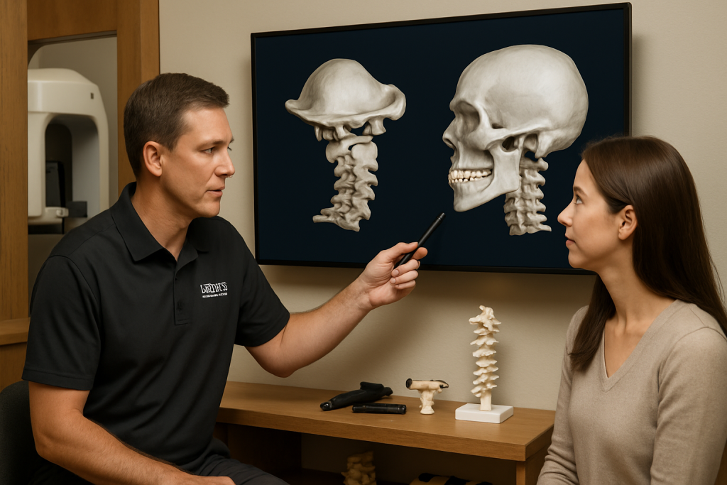
In the world of upper cervical chiropractic care, precision is everything. At Krieg Chiropractic Center in Missoula, Montana, Dr. Kevin Krieg utilizes state-of-the-art advanced imaging technology to achieve unparalleled diagnostic accuracy in Blair upper cervical technique. This commitment to precision diagnosis through advanced imaging sets the foundation for the most effective, gentle, and long-lasting upper cervical corrections possible.
Understanding how advanced imaging transforms upper cervical diagnosis can help you appreciate why this technology is essential for optimal treatment outcomes and why choosing a practitioner with these capabilities makes a significant difference in your care experience and results.
The Evolution of Upper Cervical Diagnosis
Traditional Diagnostic Limitations
Historical Challenges in Upper Cervical Assessment: Before advanced imaging technology, upper cervical practitioners faced significant diagnostic limitations:
Standard X-ray limitations – basic two-dimensional views often missed subtle misalignmentsEstimation-based analysis – practitioners relied heavily on visual interpretation and estimationLimited measurement precision – manual measurement tools introduced significant margin of errorIncomplete anatomical visualization – inability to see complex three-dimensional relationships
Consequences of Imprecise Diagnosis:
Inaccurate correction vectors leading to less effective adjustmentsMultiple adjustment attempts to achieve proper correctionLonger treatment times due to trial-and-error approachesInconsistent outcomes varying significantly between practitioners
The Advanced Imaging Revolution
Modern upper cervical care has been transformed by sophisticated imaging technology that provides unprecedented diagnostic precision:
Technological Advances:
Digital radiography offering superior image quality and analysis capabilitiesCone beam computed tomography (CBCT) providing three-dimensional visualizationComputer-assisted analysis eliminating human measurement errorVector analysis software calculating precise correction angles and forces
Clinical Impact:
Submillimeter accuracy in measuring atlas and axis position Individualized correction protocols based on specific anatomical findingsPredictable treatment outcomes through precise diagnostic informationReduced treatment time with more effective initial corrections
Advanced Imaging Technologies in Upper Cervical Care
Digital Radiography Systems
Technical Specifications: Modern digital X-ray systems used in upper cervical diagnosis offer:
Enhanced resolution – up to 10 times greater detail than traditional filmImmediate image availability – no waiting for film developmentDigital manipulation capabilities – adjusting contrast, brightness, and magnificationReduced radiation exposure – up to 90% less radiation than conventional X-rays
Clinical Applications:
Static structural analysis – precise measurement of bone position and alignmentComparative studies – before and after treatment documentationProgress monitoring – tracking improvements over timePatient education – visual demonstration of problems and improvements
Blair Technique Integration: Digital radiography is essential for Blair technique precision:
Nasium X-ray views – specialized anterior-to-posterior images unique to Blair analysisLateral cervical films – detailed side views showing atlas-axis relationshipsVertex views – top-down perspective revealing rotational componentsBase posterior views – specialized angles showing specific atlas positioning
Cone Beam Computed Tomography (CBCT)
Revolutionary Three-Dimensional Imaging: CBCT technology represents the pinnacle of upper cervical diagnostic imaging:
Technical Advantages:
3D reconstruction – complete visualization of atlas-axis complex from any angleCross-sectional analysis – examination of structures in multiple planes simultaneouslyBone density assessment – evaluation of structural integrity and healthSoft tissue visualization – limited assessment of ligaments and muscle relationships
Clinical Benefits:
Complex case analysis – essential for challenging diagnostic situationsSurgical planning – when conservative care isn’t sufficientResearch applications – advancing understanding of upper cervical biomechanicsTeaching tool – exceptional educational value for patient understanding
Blair Technique Enhancement: CBCT imaging elevates Blair analysis to new levels:
Vector precision – three-dimensional correction angle calculationAnatomical variation detection – identifying unique individual structural differencesJoint space analysis – detailed assessment of atlantooccipital and atlantoaxial jointsRotational component measurement – precise quantification of complex misalignment patterns
Computer-Assisted Analysis Systems
Digital Measurement Precision: Advanced software systems eliminate human error in diagnostic analysis:
Analysis Capabilities:
Automated landmark identification – computer recognition of anatomical reference pointsPrecision measurement – submillimeter accuracy in distance and angle calculationsStatistical analysis – comparison to normal population parametersTrend tracking – longitudinal analysis of treatment progress
Blair Technique Software: Specialized programs designed specifically for Blair analysis:
Vector calculation algorithms – precise determination of correction directionsAnatomical modeling – three-dimensional representation of patient-specific anatomyTreatment planning – optimal adjustment strategy developmentOutcome prediction – expected correction results based on analysis
The Blair Technique Diagnostic Protocol
Comprehensive Imaging Sequence
Initial Assessment Imaging: At Krieg Chiropractic Center, Dr. Krieg follows a systematic approach to upper cervical imaging:
Pre-Adjustment Series:
Nasium view – anterior-to-posterior image showing atlas lateral displacementLateral cervical – side view revealing anterior-posterior positioningVertex view – specialized top-down image showing rotational componentsBase posterior – unique Blair view demonstrating specific atlas orientation
Advanced Analysis When Indicated:
CBCT scanning for complex cases requiring three-dimensional analysisFlexion-extension studies when dynamic instability is suspectedComparative views with previous imaging when availableStress radiographs in cases of suspected ligamentous injury
Precision Measurement Protocols
Atlas Position Analysis: Advanced imaging allows precise measurement of atlas displacement in all three planes:
Lateral Displacement (Laterality):
Measurement parameters – distance of atlas lateral mass displacementNormal ranges – typically less than 2mm from midlineClinical significance – lateral displacement patterns and neurological effectsCorrection vectors – precise angles for optimal adjustment delivery
Anterior-Posterior Positioning:
Atlas rotation – measurement of forward or backward tiltingJoint space analysis – assessment of atlantooccipital and atlantoaxial gapsLigamentous stress – evaluation of structural strain patternsCompensatory changes – identification of secondary adaptations
Rotational Components:
Axial rotation – measurement of atlas rotation around vertical axisComplex patterns – identification of multi-planar misalignment combinationsCompensation assessment – evaluation of C2 and lower cervical adaptationsFunctional implications – correlation between rotation and symptom patterns
Axis Evaluation Protocols
C2 Vertebra Analysis :
Odontoid Process Assessment:
Dens positioning – evaluation of odontoid process alignmentJoint integrity – assessment of atlantoaxial joint stabilityRotational capacity – measurement of available movement rangesPathological changes – identification of degenerative or traumatic alterations
Axis Body Evaluation:
Vertebral body alignment – assessment of C2 position relative to C3Spinous process deviation – measurement of rotational componentsFacet joint analysis – evaluation of bilateral C2-C3 joint relationshipsNeural foraminal assessment – examination of nerve root exit pathways
Clinical Applications of Precision Diagnosis
Treatment Planning Enhancement
Individualized Correction Strategies: Advanced imaging enables Dr. Krieg to develop highly specific treatment protocols:
Vector Analysis:
Precise angles – exact directions for adjustment force applicationForce magnitude – optimal pressure requirements for correctionContact points – specific anatomical locations for adjustment deliverySafety considerations – identification of anatomical variations affecting treatment
Correction Sequencing:
Primary dysfunction – identification of the most significant misalignmentSecondary compensations – recognition of adaptive changes requiring attentionTreatment phases – logical progression of correction strategiesOutcome prediction – expected timeline and response patterns
Progress Monitoring and Documentation
Objective Outcome Measurement: Advanced imaging provides concrete evidence of treatment effectiveness:
Post-Adjustment Analysis:
Immediate correction verification – confirmation of successful adjustment deliveryAlignment improvement – quantification of structural changes achievedStability assessment – evaluation of correction maintenance over timeTreatment refinement – adjustments to technique based on response patterns
Long-term Tracking:
Structural improvements – documentation of progressive alignment enhancementDegenerative changes – monitoring of joint health and preservationFunctional correlation – relationship between structural and symptomatic improvementsMaintenance protocols – development of individualized care schedules
Challenging Case Management
Complex Diagnostic Scenarios: Advanced imaging is essential for difficult upper cervical cases:
Post-Traumatic Conditions:
Accident-related injuries – comprehensive assessment of trauma-induced changesLigamentous damage – evaluation of structural integrity following injuryInflammatory responses – identification of tissue changes affecting treatmentHealing progression – monitoring of recovery patterns over time
Degenerative Conditions:
Arthritic changes – assessment of joint degeneration impact on treatmentBone spur formation – identification of bony growths affecting adjustmentDisc degeneration – evaluation of intervertebral disc healthAge-related changes – consideration of normal aging processes in treatment planning
Congenital Variations:
Anatomical anomalies – identification of unusual bone formationsDevelopmental differences – recognition of growth-related variationsFamilial patterns – understanding hereditary influences on structureTreatment modifications – adaptation of techniques for unique anatomy
Patient Benefits of Advanced Imaging Diagnosis
Enhanced Treatment Precision
Improved Outcomes: Patients at Krieg Chiropractic Center benefit significantly from advanced imaging diagnosis:
More Effective Adjustments:
First-time success – higher likelihood of achieving correction on initial attemptReduced treatment visits – fewer adjustments needed due to precisionLonger-lasting corrections – better stability through accurate initial positioningImproved symptom resolution – more effective addressing of underlying problems
Reduced Discomfort:
Gentler adjustments – precise positioning eliminates need for forceful manipulationMinimal tissue trauma – accurate technique reduces surrounding tissue stressFaster recovery – less inflammation and soreness following treatmentEnhanced comfort – improved patient experience throughout treatment process
Safety Enhancement
Risk Minimization: Advanced imaging significantly improves treatment safety:
Anatomical Awareness:
Vascular considerations – identification of vertebral artery variationsBone quality assessment – evaluation of structural integrity before treatmentLigamentous status – understanding of joint stability and contraindicationsPathological identification – recognition of conditions requiring modified approaches
Treatment Contraindications:
Fracture detection – identification of bone injuries requiring alternative careTumor recognition – discovery of pathological processes affecting treatmentInfection identification – detection of inflammatory conditionsInstability assessment – recognition of excessive joint mobility requiring stabilization
Patient Education and Understanding
Visual Learning: Advanced imaging serves as powerful educational tools:
Problem Demonstration:
Clear visualization – patients can see their specific misalignment patternsBefore and after comparisons – dramatic demonstration of treatment effectivenessEducational value – enhanced understanding of anatomy and dysfunctionMotivation enhancement – visual proof of need for and benefit from treatment
Treatment Explanation:
Procedure clarification – showing exactly what will be correctedExpected outcomes – realistic preview of anticipated improvementsHome care understanding – visual reinforcement of care instructionsLong-term planning – demonstration of maintenance needs and benefits
Technology Integration at Krieg Chiropractic Center
State-of-the-Art Equipment
Digital Radiography System: Dr. Krieg utilizes the latest digital X-ray technology:
Equipment Features:
High-resolution sensors – maximum image quality for detailed analysisInstant image processing – immediate availability for analysis and consultationDose optimization – minimum radiation exposure while maintaining image qualityErgonomic design – comfortable positioning for patients during imaging
Clinical Integration:
Immediate analysis – on-the-spot evaluation and treatment planningPatient consultation – real-time discussion of findings and treatment optionsTreatment verification – immediate confirmation of successful correctionsProgress documentation – systematic tracking of improvement over time
Advanced Analysis Software
Blair Technique Software: Specialized computer programs designed specifically for Blair analysis:
Measurement Capabilities:
Automated calculations – elimination of human measurement errorStatistical comparisons – assessment relative to normal population parametersTrend analysis – tracking of changes over multiple visitsReport generation – professional documentation for patient records and referrals
Clinical Benefits:
Consistency – standardized analysis protocols ensuring reliable resultsAccuracy – submillimeter precision in measurements and calculationsEfficiency – rapid analysis allowing more time for patient careDocumentation – comprehensive records supporting treatment decisions
Quality Assurance Protocols
Equipment Maintenance: Ensuring optimal imaging quality through systematic maintenance:
Regular Calibration:
Daily quality checks – verification of equipment performance parametersMonthly calibration – professional adjustment of imaging systemsAnnual inspections – comprehensive evaluation by qualified techniciansImmediate repairs – prompt attention to any equipment issues
Image Quality Control:
Exposure optimization – balancing image quality with radiation dosePositioning accuracy – ensuring consistent and reproducible patient positioningAnalysis verification – double-checking critical measurements and calculationsContinuing education – ongoing training in latest imaging techniques and technology
The Future of Advanced Imaging in Upper Cervical Care
Emerging Technologies
Artificial Intelligence Integration: The future of upper cervical imaging includes AI-enhanced analysis:
Machine Learning Applications:
Pattern recognition – AI identification of subtle misalignment patternsOutcome prediction – computer modeling of treatment response probabilitiesTreatment optimization – AI-assisted development of correction strategiesQuality enhancement – automated image improvement and analysis
Advanced Imaging Modalities:
3D motion analysis – dynamic assessment of upper cervical movement patternsMRI integration – enhanced soft tissue visualization capabilitiesUltrasound applications – real-time assessment of tissue changesBiomarker analysis – correlation of imaging findings with physiological markers
Research and Development
Ongoing Studies: Research continues to advance upper cervical imaging capabilities:
Validation Studies:
Measurement accuracy – verification of imaging precision and reliabilityClinical correlation – relationship between imaging findings and patient outcomesTechnology comparison – evaluation of different imaging modalitiesCost-effectiveness – analysis of imaging benefits relative to costs
Innovation Development:
Portable imaging – development of mobile upper cervical imaging systemsRadiation reduction – continued advancement in dose optimizationAnalysis automation – further development of computer-assisted analysisIntegration platforms – comprehensive systems combining multiple imaging modalities
Choosing Advanced Imaging Upper Cervical Care
What to Look for in a Practice
Essential Capabilities: When seeking upper cervical care, look for practices with advanced imaging capabilities:
Technology Requirements:
Digital radiography – minimum standard for precise upper cervical analysisSpecialized software – computer-assisted analysis for maximum accuracyRegular updates – commitment to maintaining current technology standardsQuality assurance – systematic protocols ensuring optimal imaging quality
Practitioner Qualifications:
Specialized training – specific education in advanced imaging interpretationCertification – credentials in upper cervical technique requiring imaging expertiseExperience – extensive background in complex case analysis and managementContinuing education – ongoing learning in imaging advances and applications
The Krieg Chiropractic Center Advantage
Comprehensive Imaging Services: Dr. Kevin Krieg’s practice offers the complete range of advanced imaging capabilities:
Technology Portfolio:
State-of-the-art digital radiography – highest quality upper cervical imagingAdvanced analysis software – computer-assisted precision measurementComprehensive protocols – systematic approach to diagnostic imagingRegular upgrades – commitment to maintaining cutting-edge capabilities
Clinical Expertise:
Blair technique certification – specialized training in precision imaging analysis20+ years experience – extensive background in challenging upper cervical casesResearch involvement – participation in advancing upper cervical imaging scienceEducational commitment – teaching and mentoring other practitioners
Patient-Centered Approach:
Detailed explanation – thorough education about imaging findingsVisual demonstration – clear presentation of problems and solutionsShared decision-making – collaborative treatment planning based on imaging resultsOngoing monitoring – systematic tracking of progress through follow-up imaging
Conclusion: Precision as the Foundation of Excellence
Advanced imaging technology has revolutionized upper cervical chiropractic care , transforming it from an art based on clinical experience to a precision science based on objective measurement and analysis. At Krieg Chiropractic Center in Missoula, Montana, Dr. Kevin Krieg’s commitment to utilizing the most advanced imaging technology available ensures that patients receive the most precise, effective, and safe upper cervical care possible.
The integration of digital radiography, computer-assisted analysis, and specialized Blair technique software creates an unprecedented level of diagnostic accuracy that directly translates into superior treatment outcomes. This precision approach means fewer visits, longer-lasting corrections, and more predictable results for patients seeking relief from upper cervical dysfunction.
The investment in advanced imaging technology reflects a commitment to excellence that extends beyond just having the latest equipment. It represents a philosophy that recognizes the critical importance of accurate diagnosis as the foundation for effective treatment. When your atlas and axis vertebrae require correction, precision isn’t just beneficial – it’s essential.
Understanding the role of advanced imaging in upper cervical care empowers you to make informed decisions about your healthcare and choose practitioners who have invested in the technology and training necessary to provide the highest quality care available.
Ready to experience the precision of advanced imaging diagnosis? Contact Krieg Chiropractic Center at (406) 541-8888 to schedule your comprehensive upper cervical evaluation and discover how state-of-the-art imaging technology can provide the accurate diagnosis essential for your optimal health outcomes.